RadioMatrix™
RadioMatrix™ for Radiopaque Medical Models
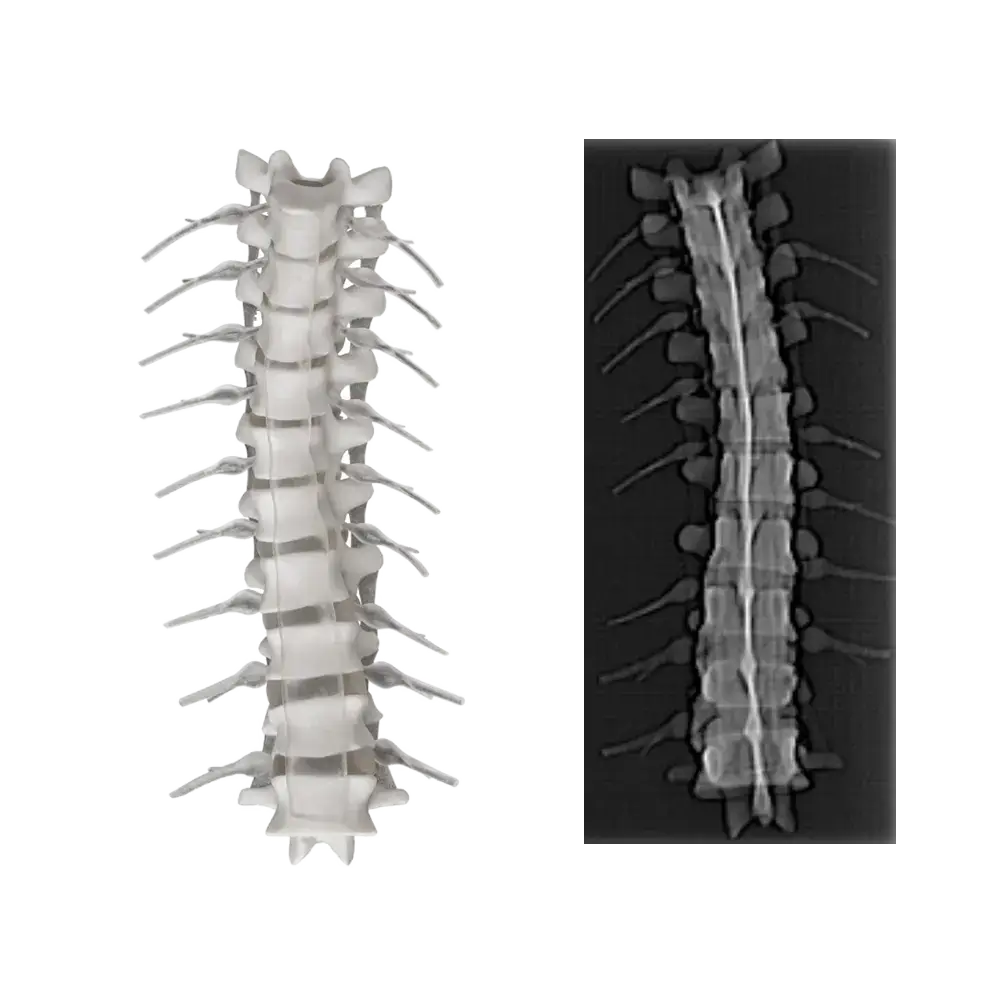
RadioMatrix™ is a radiopaque 3D printing material used to create models that are visible under standard medical imaging methods. It enables the production of anatomically accurate parts with density levels that mimic human tissue, supporting real-world medical applications from surgical simulation to device validation.
CT and X-Ray Compatibility
RadioMatrix™ produces consistent, high-contrast parts that appear clearly in CT and X-ray scans. This makes it suitable for testing medical devices, planning surgeries, and educating practitioners using visual feedback that matches clinical imaging environments.
Mimics Tissue Densities
Engineered to simulate a wide range of Hounsfield Unit (HU) values, this material allows you to replicate specific tissue types within a single part or across a full-body region. It enables realistic medical modeling with traceable, repeatable imaging results.
Supports Clinical Confidence
By delivering lifelike imaging and reliable mechanical performance, RadioMatrix™ helps reduce guesswork during planning and supports accurate rehearsals before procedures. It also enhances collaboration between clinical teams and device engineers.
Efficient for Research and Training
This material supports the development of training aids, educational tools, and device testbeds that replicate real-life scenarios. It offers medical teams and innovators a low-risk environment to simulate, test, and refine their approach with scan-visible results.